Smooth Muscle Diagram : Overview Of The Muscular System Boundless Anatomy And Physiology : Smooth muscle is a type of muscle tissue which is used by various systems to apply pressure to vessels and organs.. In skeletal muscle, a single type of somatic nervous system traverses to muscle, where it stimulates organelle in the muscle cells in order to release calcium. Smooth muscle is a type of muscle tissue which is used by various systems to apply pressure to vessels and organs. 1024x840 draw a labelled diagram of a smooth muscle diagram of smooth. The smooth muscles perform the functions in the contrast of other types of muscles. Its wavelike movements propel things through the bodily system, such as food through.
This figure shows the structure of the muscle fibers. Smooth muscle diagram | quizlet. Note that the smooth muscle cells are arranged in layers that are orthagonal to each other. Smooth muscle cells are under control of the autonomous nervous system. The pupillary sphincter muscle in your eye is a smooth muscle that shrinks the size of your.
How Many Muscles Are In The Human Body Plus A Diagram from post.healthline.com These cells have fibers of actin and myosin which run through the cell and are supported by a framework of other proteins. Back muscle chart 12 photos of the back muscle chart back muscle diagram human body, back muscle diagram pain, back muscle groups diagram, back muscle workout diagram, lower back muscle chart, human muscles, back muscle diagram human body, back muscle diagram pain, back muscle groups diagram, back muscle workout diagram. In a test of biology a figure of smooth muscle labeled as a, b, c and d for different parts of the muscles. Smooth muscle contracts under certain stimuli as atp is freed. Note that the smooth muscle cells are arranged in layers that are orthagonal to each other. It is the weakest type of muscle but smooth muscles in the gastrointestinal or gi tract control digestion. Vascular smooth muscle refers to the particular type of smooth muscle found within, and composing the majority of the wall of blood vessels. It constitutes much of the musculature of
In addition, the contractile state of smooth muscle is controlled by hormones, autocrine/paracrine agents, and other local chemical signals.
Its wavelike movements propel things through the bodily system, such as food through. Beta 2 receptors are also on small coronary arterioles thus increasing hormonally induced blood flow within the musculature of the heart. In a test of biology a figure of smooth muscle labeled as a, b, c and d for different parts of the muscles. Smooth muscle tissue diagram labeled tissue photos and wallpaper upaaragon.co. This is just a diagram of how the human muscle looks under all the tissue and skin. In addition, the contractile state of smooth muscle is controlled by hormones, autocrine/paracrine agents, and other local chemical signals. Arteries have thick walls due to smooth muscle cells, which help them carry blood away from the heart to every part of. The movement is caused when these muscles contract and relax. Smooth muscle diagram | quizlet. It is the main muscle of respiration. The calcium is the cause of protein to detach from the actin and myosin fastly binds with the opening of actin. This figure shows the structure of the muscle fibers. Smooth muscle makes up the walls of hollow organs, respiratory passageways, and blood vessels.
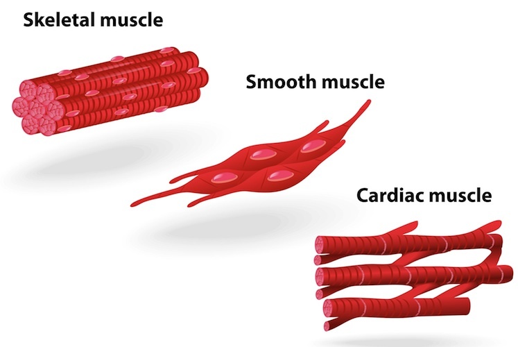
It is the pen diagram of skeletal, smooth and cardiac muscle for class 10, 11 and 12. 1024x840 draw a labelled diagram of a smooth muscle diagram of smooth. Smooth muscle is a type of muscle tissue which is used by various systems to apply pressure to vessels and organs. Smooth muscle is composed of sheets or strands of smooth muscle cells. Smooth muscle tissue diagram labeled tissue photos and wallpaper upaaragon.co.
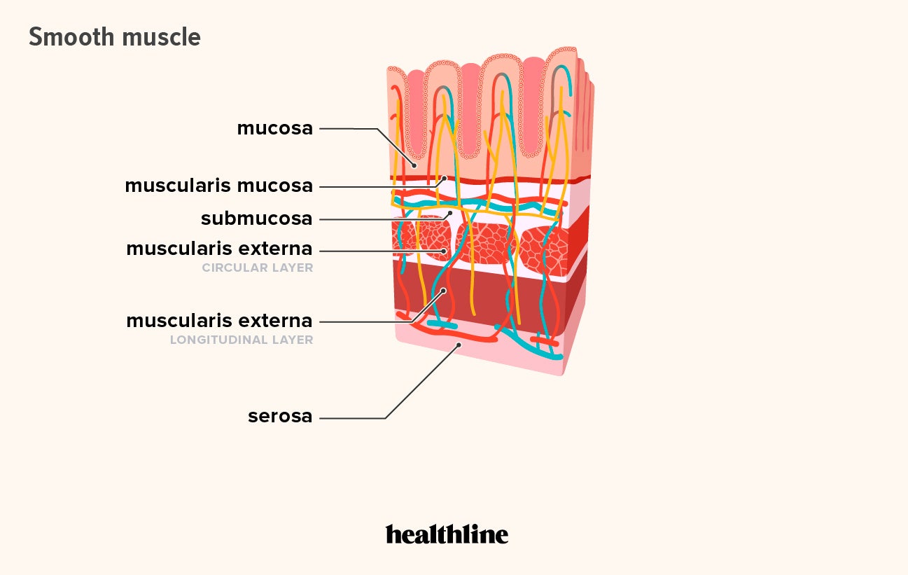
Muscle Structure Muscle Under The Microscope Science Learning Hub from static.sciencelearn.org.nz It is the pen diagram of skeletal, smooth and cardiac muscle for class 10, 11 and 12. 1024x840 draw a labelled diagram of a smooth muscle diagram of smooth. Ncert cbse class ix science. The movement is caused when these muscles contract and relax. It is layered in a distinctive pattern of circular layers. Smooth muscle, muscle that shows no cross stripes under microscopic magnification. Smooth muscle makes up the walls of hollow organs, respiratory passageways, and blood vessels. Smooth muscle diagram | quizlet.
Smooth muscle tissue is also known as visceral muscle tissue.
Smooth muscle is composed of sheets or strands of smooth muscle cells. This is just a diagram of how the human muscle looks under all the tissue and skin. Vascular smooth muscle refers to the particular type of smooth muscle found within, and composing the majority of the wall of blood vessels. In skeletal muscle, a single type of somatic nervous system traverses to muscle, where it stimulates organelle in the muscle cells in order to release calcium. The cardiac muscle is only found in the heart wall. Gap junctions between cells allows coordination of contraction. Vascular smooth muscle is the type of smooth muscle that makes up most of the walls of blood vessels. 1024x840 draw a labelled diagram of a smooth muscle diagram of smooth. Smooth muscle is a type of muscle tissue which is used by various systems to apply pressure to vessels and organs. Beta 2 receptors are also on small coronary arterioles thus increasing hormonally induced blood flow within the musculature of the heart. Smooth muscle tissue is also known as visceral muscle tissue. Smooth muscle tissue, unlike striated muscle, contracts slowly and automatically. Smooth muscle cells lack the striated banding pattern found in cardiac and skeletal muscle, and they receive neural innervation from the autonomic nervous system.
Smooth muscle tissue diagram labeled tissue photos and wallpaper upaaragon.co. Smooth muscle is composed of sheets or strands of smooth muscle cells. In arteries, smooth muscle movements maintain the arteries' diameter. It is layered in a distinctive pattern of circular layers. This figure shows the structure of the muscle fibers.
Resp 8 Airway Smooth Muscle Diagram Quizlet from o.quizlet.com The calcium is the cause of protein to detach from the actin and myosin fastly binds with the opening of actin. Smooth muscle makes up the walls of hollow organs, respiratory passageways, and blood vessels. Its wavelike movements propel things through the bodily system, such as food through. This figure shows the structure of the muscle fibers. The smooth muscles perform the functions in the contrast of other types of muscles. Note that the smooth muscle cells are arranged in layers that are orthagonal to each other. It is the pen diagram of skeletal, smooth and cardiac muscle for class 10, 11 and 12. Smooth muscles in a woman's uterus (or womb) help to push babies out of the body during childbirth.
• smooth muscles respond to stretch only briefly, and then adapts to its new length • the new length however, retains its original _____ seconds or minutes after it has been elongated or shortened (e.g.
The cardiac muscle is only found in the heart wall. Smooth muscle tissue, unlike striated muscle, contracts slowly and automatically. It is layered in a distinctive pattern of circular layers. How to draw labelled diagram of smooth muscle fibre step by stephello friends in this video i tell you about how to draw smooth muscle fibre step by s. Smooth muscles exhibits a phenomenon called _____ in which: Smooth muscles exhibits a phenomenon called _____ in which: Smooth muscle is composed of sheets or strands of smooth muscle cells. In skeletal muscle, a single type of somatic nervous system traverses to muscle, where it stimulates organelle in the muscle cells in order to release calcium. Arteries have thick walls due to smooth muscle cells, which help them carry blood away from the heart to every part of. Smooth muscle contracts under certain stimuli as atp is freed. Back muscle chart 12 photos of the back muscle chart back muscle diagram human body, back muscle diagram pain, back muscle groups diagram, back muscle workout diagram, lower back muscle chart, human muscles, back muscle diagram human body, back muscle diagram pain, back muscle groups diagram, back muscle workout diagram. → the cells of cardiac muscles are cylindrical, branched, and uninucleate. This is just a diagram of how the human muscle looks under all the tissue and skin.